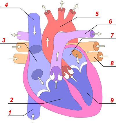
45 structure of the heart without labels
diagram of heart without labels 13 Best Images of Hip Anatomy Of The Worksheet - Sunflower Anatomy. 11 Pics about 13 Best Images of Hip Anatomy Of The Worksheet - Sunflower Anatomy : u414adad: heart diagram without labels, Congestive Heart Failure: The Essence of Heart Failure Course | CEUfast and also 13 Best Images of Hip Anatomy Of The Worksheet - Sunflower Anatomy. ear diagram without labels 11 Best Images Of Skeleton Labeling Worksheet - Skeleton Bones bones pelvic girdle diagram unlabeled labeling worksheet skeleton worksheeto via Drag Each Label To The Appropriate Location On This Diagram Of The atkinsjewelry.blogspot.com respiratory system label diagram human appropriate drag each location quiz
Human Heart Diagram Without Labels | Human heart diagram, Heart diagram ... This exhibit depicts the anatomy of the inferior skull including: the foramen magnum, occipital condyles, mastoid process, styloid process, mandibular fossa, palatine bone, sphenoid bone, carotid canal, and the jugular fossa. E Emely cale Pish Posh
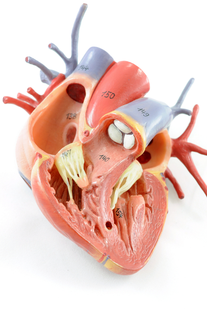
Structure of the heart without labels
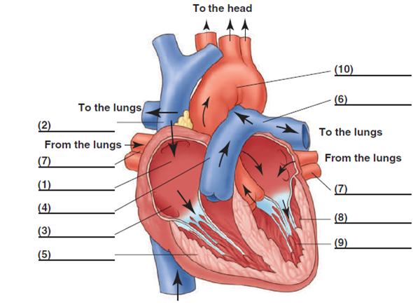
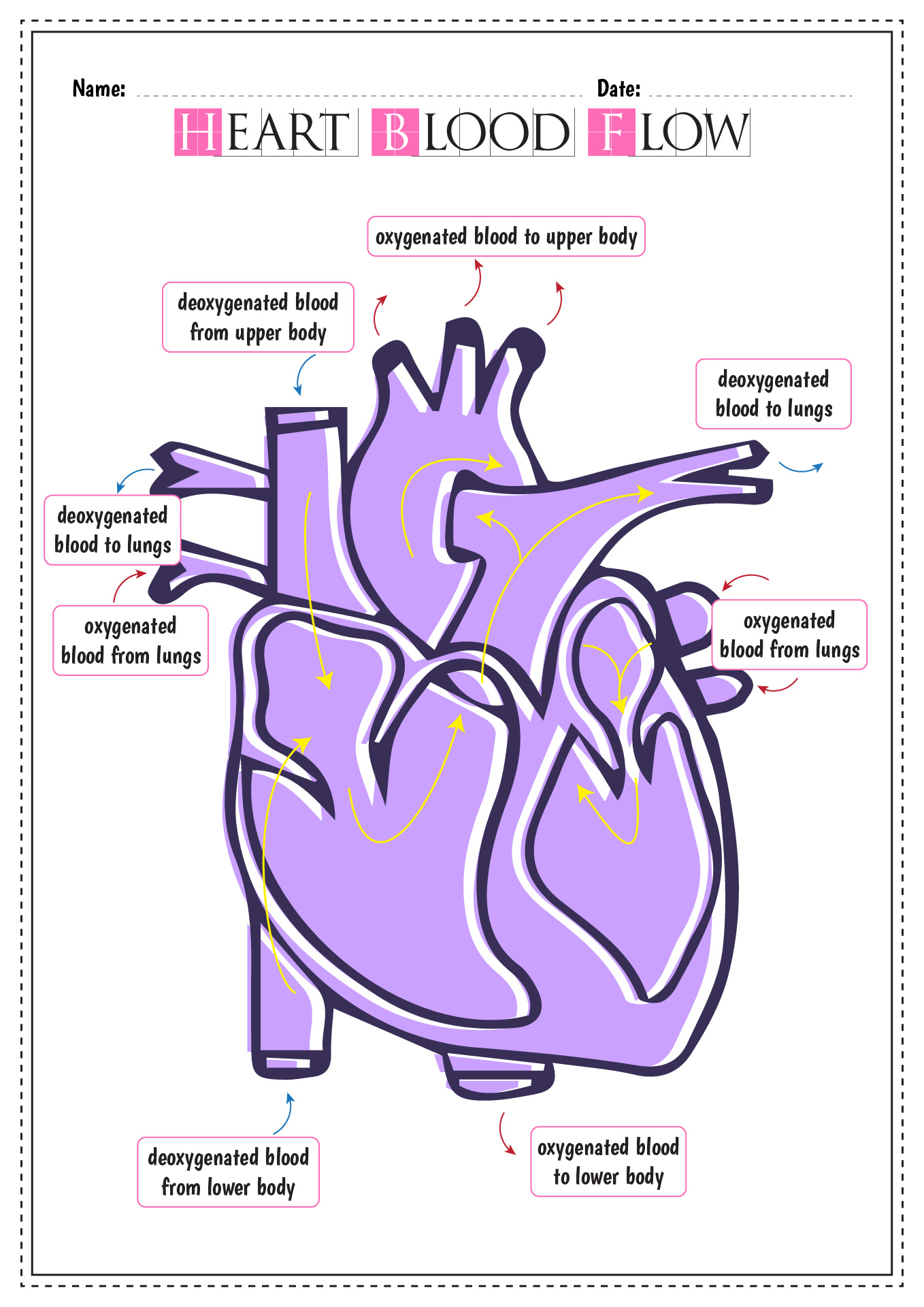
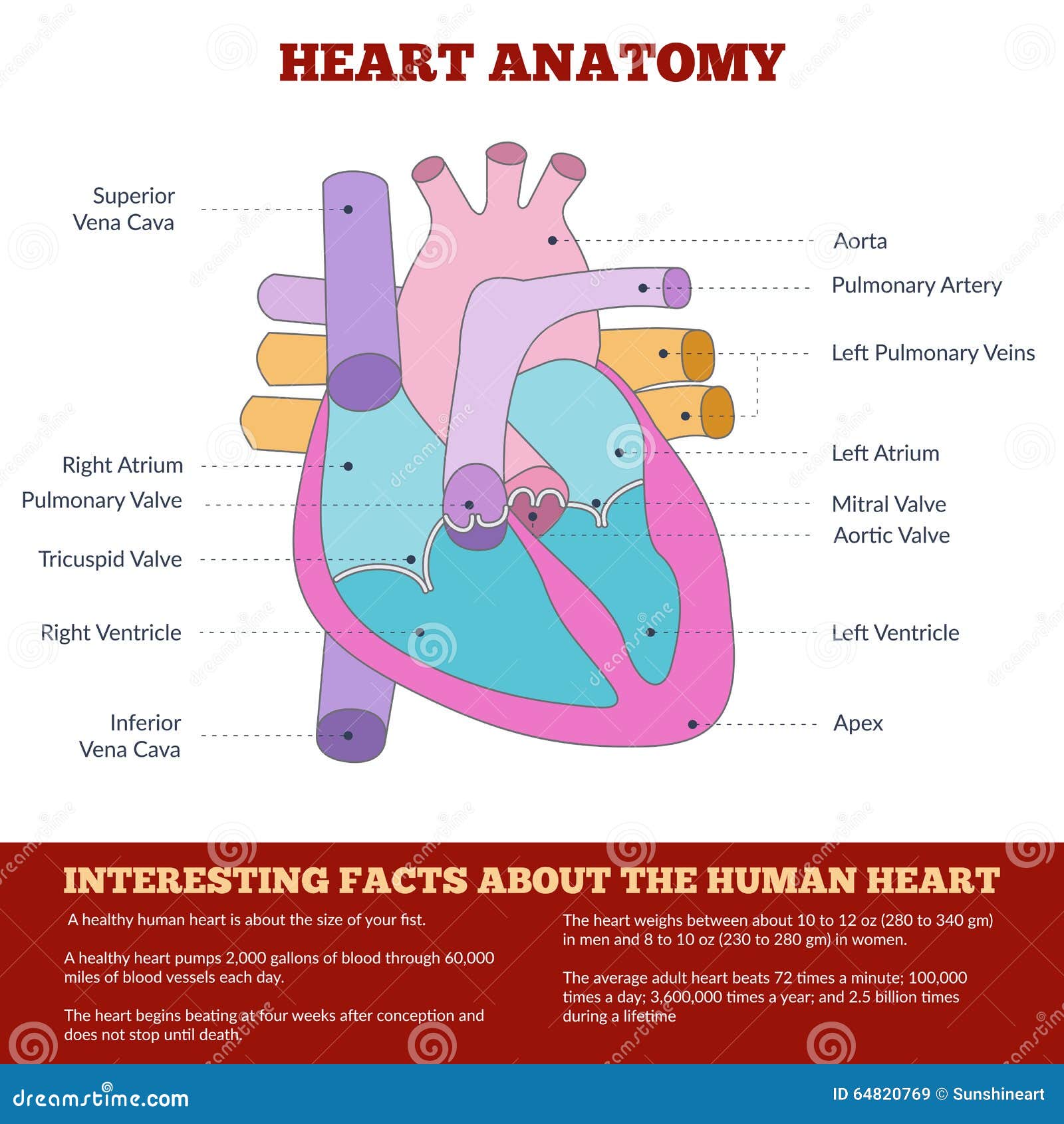
Human Heart - Diagram and Anatomy of the Heart - Innerbody Because the heart points to the left, about 2/3 of the heart's mass is found on the left side of the body and the other 1/3 is on the right. Anatomy of the Heart Pericardium. The heart sits within a fluid-filled cavity called the pericardial cavity. The walls and lining of the pericardial cavity are a special membrane known as the pericardium. circulatory system worksheet without labels - Google Search | Heart ... The areas of the heart with MORE oxygen are labeled with an "R". Students will color these areas RED. The areas of the heart with LESS oxygen are labeled with a "B". Students will color these areas BLUE. This diagram is a excellent way to visually represent the direction of blood flow through the heart. Human Heart Diagram Without Labels | Human heart diagram, Heart diagram ... Arrows represent the flow of blood. Labels include the right and left common carotid arteries, pulmonary arteries, pulmonary veins, and lungs and the right and left atrium and ventricles, the left subclavian artery, aorta, jugular veins, thoracic aorta, inferior and superior vena cava and capillaries. M varaprasad miriyala Health and Body
Structure of the heart without labels. heart diagram without labels Label The Heart Worksheets (SB6634) - SparkleBox . heart label worksheets diagram human anatomy sparklebox science body ks2 labeling physiology nursing system circulatory diagrams preschool study. Heart anatomy interior labels section cross blood vessels. Free blank heart diagram, download free blank heart diagram png images. heart diagram without labels heart diagram without labels 13+ heart diagram templates - sample, example, format download. Heart label worksheets diagram human anatomy sparklebox science body ks2 labeling physiology nursing system circulatory diagrams study. Heart diagram label parts template sheet format sample example student templates response blood A Labeled Diagram of the Human Heart You Really Need to See The human heart, comprises four chambers: right atrium, left atrium, right ventricle and left ventricle. The two upper chambers are called the left and the right atria, and the two lower chambers are known as the left and the right ventricles. The two atria and ventricles are separated from each other by a muscle wall called 'septum'. Heart Diagram with Labels and Detailed Explanation - BYJUS Diagram of Heart. The human heart is the most crucial organ of the human body. It pumps blood from the heart to different parts of the body and back to the heart. The most common heart attack symptoms or warning signs are chest pain, breathlessness, nausea, sweating etc. The diagram of heart is beneficial for Class 10 and 12 and is frequently ...
Anatomy of the Heart - Medical Animation - YouTube This medical animation demonstrates the anatomy of the human heart, while explaining how the cardiovascular system functions. Explore more of our medical ani... The Heart | Boundless Anatomy and Physiology | | Course Hero The myocardium is the muscle tissue of the heart, composed of cardiac muscle cells called cardiomyocytes that receive nervous stimulation from the sinoatrial (SA) and atrioventricular (AV nodes via the Purkinje fibers. Cardiomyocytes are shorter than skeletal myocytes, and contain fewer nuclei. Cardiac muscle is striated. heart without label Vector Winner Golden Label 458912 Vector Art At Vecteezy . winner vector golden label vecteezy. Netizens Have Said TXT YeonJun Looks Like This Former Wanna One Member . txt yeonjun member wanna netizens former said looks kpopmap enregistrée depuis. Normal Chest Axial Anatomy - Plain And Labeled Sections - YouTube Figuring Out Cardiac Anatomy: Your Heart - dummies In fact, your heart is situated slightly to the left of center in your chest. Figure 1: Anterior view of the heart. A thick layer of muscle tissue and a protective membrane that folds into two layers, called the pericardium or pericardial membranes, surround the heart. The heart itself is a well-organized grouping of hollow spaces.
Heart: Anatomy and Function - Cleveland Clinic Heart. Your heart is the main organ of your cardiovascular system, a network of blood vessels that pumps blood throughout your body. It also works with other body systems to control your heart rate and blood pressure. Your family history, personal health history and lifestyle all affect how well your heart works. Appointments 800.659.7822. Human Heart (Anatomy): Diagram, Function, Chambers, Location in Body Human Heart (Anatomy): Diagram, Function, Chambers, Location in Body The right atrium receives blood from the veins and pumps it to the right ventricle. The right ventricle receives blood from the... Human Heart Diagram Without Labels | Human heart diagram, Heart diagram ... Arrows represent the flow of blood. Labels include the right and left common carotid arteries, pulmonary arteries, pulmonary veins, and lungs and the right and left atrium and ventricles, the left subclavian artery, aorta, jugular veins, thoracic aorta, inferior and superior vena cava and capillaries. M varaprasad miriyala Health and Body circulatory system worksheet without labels - Google Search | Heart ... The areas of the heart with MORE oxygen are labeled with an "R". Students will color these areas RED. The areas of the heart with LESS oxygen are labeled with a "B". Students will color these areas BLUE. This diagram is a excellent way to visually represent the direction of blood flow through the heart.
Label Heart Structure
Human Heart - Diagram and Anatomy of the Heart - Innerbody Because the heart points to the left, about 2/3 of the heart's mass is found on the left side of the body and the other 1/3 is on the right. Anatomy of the Heart Pericardium. The heart sits within a fluid-filled cavity called the pericardial cavity. The walls and lining of the pericardial cavity are a special membrane known as the pericardium.

2.63 describe the structure of the heart and how... - phy chem bi
Solved: Label the following structures of the heart by writing ... | Chegg.com

Fish Internal Organs Vector Art Diagram Anatomy Without Labels | CartoonDealer.com #72048931
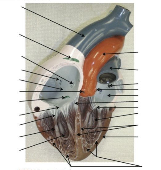
Solved: Label The Following Structures Of The Heart | Chegg.com
11 Best Images of Blank Heart Diagram Worksheet With Word Bank - Label Heart Diagram Worksheet ...
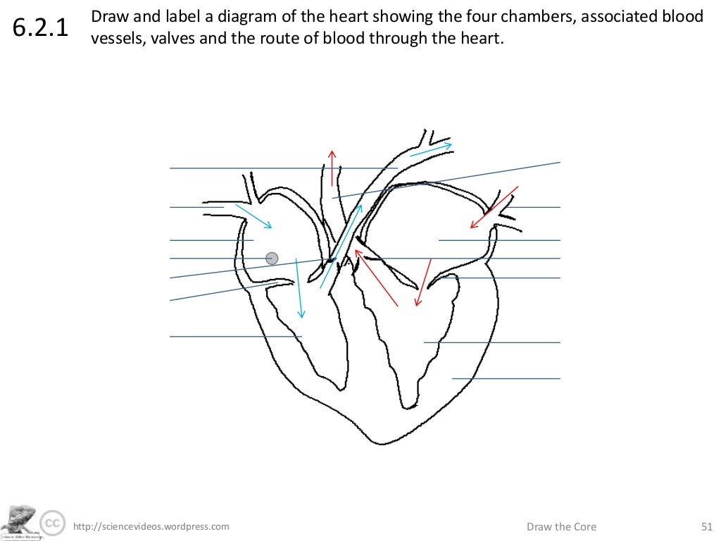
http://sciencevideos.wordpress.com Draw the Core 516.2.1

Respiratory System Without Labels Human Respiratory System | Humananatomybody | Respiratory ...
Anatomy Diagram Cartoon Vector | CartoonDealer.com #58232351


Science Stuff.: August 2010
Structure and Function of the Heart
Diagram showing anatomy of plant cell 419163 Vector Art at Vecteezy

cat muscles | Cat Muscle Dissection | Cat Dissection | Pinterest | Cats, Muscle and All in one

34 Label The Heart Diagram - Labels Database 2020
Post a Comment for "45 structure of the heart without labels"