44 parts of the eye without labels
Eye Anatomy - John Burroughs School Print a Diagram of the Eye - Click on this link and then use the browser print command to produce a diagram to use with the student tutorial. 2. Label the eye diagram. 3. Identify the parts of the eye (model). 4. Virtual sheep eye dissection. 5. Pin on Five Senses - Pinterest Description Use these simple eye diagrams to help students learn about the human eye. Three differentiated worksheets are included: 1. Write the words using a word bank 2. Cut and paste the words 3. Write the words without a word bank Labels include: eyebrow, eyelid, eyelashes, pupil, iris, and sclera. UPDATE: I've updated this product to ...
› collegehumorYouTube About Press Copyright Contact us Creators Advertise Developers Terms Privacy Policy & Safety How YouTube works Test new features
Parts of the eye without labels
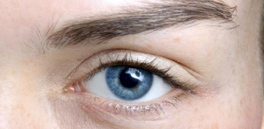
Parts of the Eye & Their Function | Robertson Optical and Optometry The iris is the area of the eye that contains the pigment which gives the eye its color. This area surrounds the pupil, and uses the dilator pupillae muscles to widen or close the pupil. This allows the eye to take in more or less light depending on how bright it is around you. If it is too bright, the iris will shrink the pupil so that they ... Diagram of eye without labels: Labelling the eye — Science Learning Hub ... Tears lubricate the eye and are made up of three layers. These three layers together are called the tear film. The mucous layer is made by the conjunctiva. The watery part of the tears is made by the lacrimal gland. The eye's lacrimal gland sits under the outside edge of the eyebrow (away from the nose) in the orbit. Parts Of The Eye Labeled Diagram Model And Their Function The parts of the eye that function include the lens, retina, vitreous body, and macula. The eyes' lenses shape the incoming light so that it can be focused on the retina, which converts this light into images that we can see. White eyes are caused by a problem with the macula or central part of the retina.
Parts of the eye without labels. en.wikipedia.org › wiki › MindMind - Wikipedia Broadly speaking, mental faculties are the various functions of the mind, or things the mind can "do". Thought is a mental act that allows humans to make sense of things in the world, and to represent and interpret them in ways that are significant, or which accord with their needs, attachments, goals, commitments, plans, ends, desires, etc. Thinking involves the symbolic or semiotic mediation ... Diagram of eye with labels Here are descriptions of some of the main parts of the eye. The cornea is the clear outer part of the eyes focusing system located at the front of the eye. The iris is the colored part of the eye that regulates the amount of light entering the eye. This is an exercise for students to label a simple blank eye diagram with the following parts. Labelling the eye — Science Learning Hub In this interactive, you can label parts of the human eye. Use your mouse or finger to hover over a box to highlight the part to be named. Drag and drop the text labels onto the boxes next to the eye diagram If you want to redo an answer, click on the box and the answer will go back to the top so you can move it to another box. › b › Sony-PlayStation-5-ConsolesSony PlayStation 5 Consoles for sale | eBay Get the best deals on Sony PlayStation 5 Consoles and upgrade your gaming setup with a new gaming console. Find the lowest prices at eBay.com. Fast & Free shipping on many items!
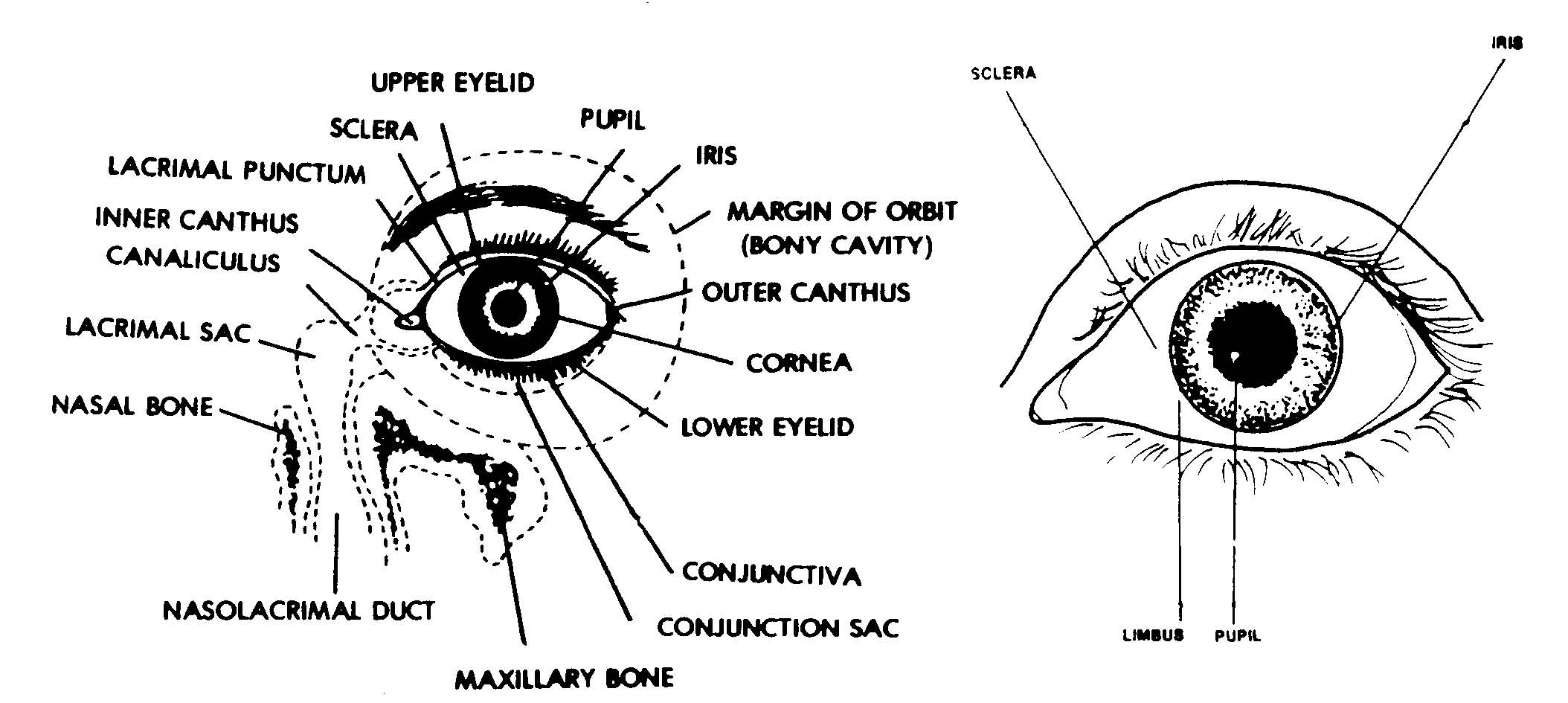
Diagram of the eye with labels Diagram of the eye with labels. The parts of the eye that are visible externally include the following-Sclera. It is a white visible portion. It is made up of dense connective tissue and protects the inner parts. It lines the sclera and is made up of stratified squamous epithelium. › latest-newsLatest News | American Cancer Society Apr 15, 2022 · We couldn’t do what we do without our volunteers and donors. Together, we’re making a difference – and you can, too. Become a volunteer, make a tax-deductible donation, or participate in a fundraising event to help us save lives. Explore Get Involved Labelling the eye — Science Learning Hub Labelling the eye Add to collection The human eye contains structures that allow it to perceive light, movement and colour differences. In this activity, students use online or paper resources to identity and label the main parts of the human eye. By the end of this activity, students should be able to: identify the main parts of the human eye Diagram of the Eye - Home - Lions Eye Institute Instructions. Click the parts of the eye to see a description for each. Hover the diagram to zoom. Iris. The iris is the coloured part of the eye which surrounds the pupil. It controls light levels inside the eye, similar to the aperture on a camera. The iris contains tiny muscles that widen and narrow the pupil size.
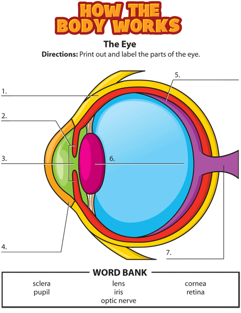
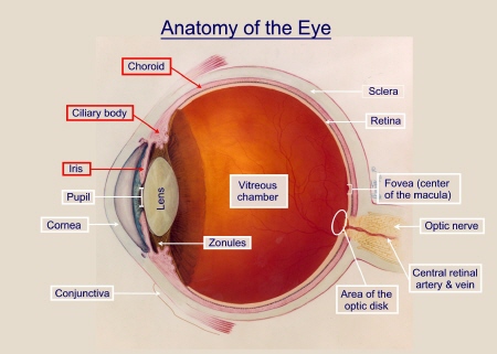
› no-longer-availableNo Longer Available - WDSU 'I need the rest of my rent money': More people turn to pawn shops as inflation continues to rise PDF Parts of the Eye Eye Diagram Handout Author: National Eye Health Education Program of the National Eye Institute, National Institutes of Health Subject: Handout illustrating parts of the eye Keywords: parts of the eye, eye diagram, vitreous gel, iris, cornea, pupil, lens, optic nerve, macula, retina Created Date: 12/16/2011 12:39:09 PM eyeball diagram to label; labels without eye hypothalamus thalamus clipart front brain label parts stem cerebellum clipground. Label The Eye . eye diagram blank human labeled anatomy label eyeball worksheet quiz drawing labels purposegames answers parts printable ear eyes activities coloring. Human Eye Diagram, How The Eye Work -15 Amazing Facts Of Eye Labeled Eye Diagram | Science Trends What you want to interpret as a major part of the human eye is somewhat up to the individual, but in general there are seven parts of the human eye: the cornea, the pupil, the iris, the lens, the vitreous humor, the retina, and the sclera. Let's take a closer look at each of these components individually. The Cornea
Label The Parts Of The Eye - ProProfs Quiz
open.umn.edu › opentextbooks › textbooksStand up, Speak out: The Practice and Ethics of Public Speaking Jun 21, 2021 · Stand up, Speak out: The Practice and Ethics of Public Speakingfeatures two key themes. First it focuses on helping students become more seasoned and polished public speakers, and second is its emphasis on ethics in communication. It is this practical approach and integrated ethical coverage that setsStand up, Speak out: The Practice and Ethics of Public Speakingapart from the other texts in ...

diagram of eye without labels Correctly Label the Eye Diagram Quiz. 11 Images about Correctly Label the Eye Diagram Quiz : Diagram Of Human Eye Without Label | MedicineBTG.com, Correctly Label the Eye Diagram Quiz and also Relapsing Polychondritis: Causes, Picture, Symptoms and Treatment.

Label The Eye Diagram - Pensandpieces
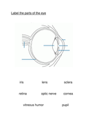
Label Parts of the Human Eye - University of Dayton Parts of the Eye Select the correct label for each part of the eye. The image is taken from above the left eye. Click on the Score button to see how you did. Incorrect answers will be marked in red.

Blank Ear Diagram | Human ear diagram, Ear anatomy, Ear diagram
Eye Anatomy: Parts of the Eye and How We See Behind the anterior chamber is the eye's iris (the colored part of the eye) and the dark hole in the middle called the pupil. Muscles in the iris dilate (widen) or constrict (narrow) the pupil to control the amount of light reaching the back of the eye. Directly behind the pupil sits the lens. The lens focuses light toward the back of the eye.

Eye anatomy: A closer look at the parts of the eye Eye anatomy: A closer look at the parts of the eye. When surveyed about the five senses — sight, hearing, taste, smell and touch — people consistently report that their eyesight is the mode of perception they value (and fear losing) most. Despite this, many people don't have a good understanding of the anatomy of the eye, how vision works ...
Vocabulary - Homeschoolers
Eye Anatomy: 16 Parts of the Eye & Their Functions The following are parts of the human eyes and their functions: 1. Conjunctiva The conjunctiva is the membrane covering the sclera (white portion of your eye). The conjunctiva also covers the interior of your eyelids. Conjunctivitis, often known as pink eye, occurs when this thin membrane becomes inflamed or swollen.

Eye Anatomy: External Parts of the Eye - Optometrists.org The external parts of the eye work together to protect the eye and all of its internal structures. The following ocular structures are located on the eye's exterior: Eyelids. The upper and lower eyelids form a moist region around the eye, and protect the surface of the eye from injury, infection, and disease.

Bimmers Garage Trading: BMW E36 E46 E39 E38 E30 E34 E32 LED ANGEL EYE
diagram of eye with labels Ear anatomy eye models lab guide structures identify biologycorner brain senses biology eyes physiology parts use answers. Peregrine diagram falcon hi res. Picture of the eye labeled elegant human eye anatomy for kids ... Eye Diagram Without Labels | Via Anatomy Pictures Gallery If… | Flickr . liver fluke. Perception: 3.1 Eye To ...

Anatomy of the eye: Quizzes and diagrams - Kenhub Found within two cavities in the skull known as the orbits, the eyes are surrounded by several supporting structures including muscles, vessels, and nerves. There are 7 bones of the orbit, two groups of muscles (intrinsic ocular and extraocular), three layers to the eyeball … and that's just the beginning. There's a lot to learn, but stay calm!

Anatomy of the Eye | Johns Hopkins Medicine Cornea. The clear, dome-shaped surface that covers the front of the eye. Iris. The colored part of the eye. The iris is partly responsible for regulating the amount of light permitted to enter the eye. Lens (also called crystalline lens). The transparent structure inside the eye that focuses light rays onto the retina. Lower eyelid.
33 Label Parts Of The Eye - Labels 2021
co.merced.ca.usMerced County, CA - Official Website | Official Website The Dos Palos Library will soon undergo a major facilities upgrade thanks to $3 million of funding secured in the state's Fiscal Year 2022-23 budget.
31 Label The Parts Of Eye - Labels For Your Ideas
the ear diagram without labels hyaloid artery anatomy ear human eyeball untpikapps unlabeled alchetron unattainable vitreous retinal excelguider arteria Dr. Yung's ENT Practice | Information: Ears ear ears diagram canal inner parts otosclerosis fistula hearing dizziness menieres treatment physical disease infection syndrome williams children wax drops

Label the Eye - PurposeGames
PDF Eye Anatomy Handout - National Eye Institute of light entering the eye. Lens: The lens is a clear part of the eye behind the iris that helps to focus light, or an image, on the retina. Macula: The macula is the small, sensitive area of the retina that gives central vision. It is located in the center of the retina. Optic nerve: The optic nerve is the largest sensory nerve of the eye.
Kids | Uveitis.org | OIUF
File:Diagram of human eye without labels.svg - Wikimedia Size of this PNG preview of this SVG file: 410 × 430 pixels. Other resolutions: 229 × 240 pixels | 458 × 480 pixels | 732 × 768 pixels | 976 × 1,024 pixels | 1,953 × 2,048 pixels. Original file (SVG file, nominally 410 × 430 pixels, file size: 277 KB) File information. Structured data.
How our eyes work | Teaching Resources
Eye Diagram With Labels and detailed description - BYJUS Iris is the coloured part of the eye and controls the amount of light entering the eye by regulating the size of the pupil. The lens is located just behind the iris. Its function is to focus the light on the retina. The optic nerve transmits electrical signals from the retina to the brain. Pupil is the opening at the centre of the iris.
Activity: Name the Parts of the Eye - American Academy of Ophthalmology
Quiz: Label The Parts Of The Eye - ProProfs How much did you get to understand about the human eye? Take up this quiz and find out! Questions and Answers. 1. A is pointing to what part of the eye? A. Cornea. B. Optic Nerve.
Post a Comment for "44 parts of the eye without labels"